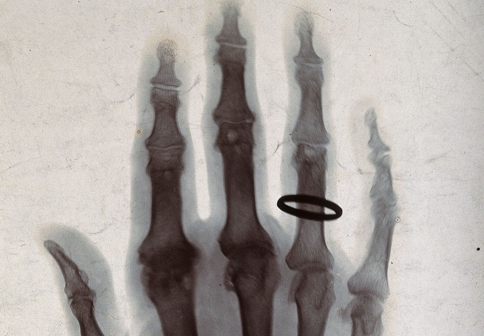
In the last days of 1895, Wilhelm Röntgen showed his wife a photograph of her own hand. The flesh had faded to a mist. The bones, and her ring, stood out as if drawn in ink.
Chapter 11 · Beyond the visible
A New Kind of Ray
Light that passes through flesh and is stopped by bone. Why bone, and not flesh? And why do doctors change the X-rays for a hand, a breast and a chest?
Look first
Layer upon layer
Among the first things Röntgen photographed with his new rays was a staircase of tin foil: one sheet, then two, then three, and on up. Each extra sheet left the plate a step paler. The rays were not stopped all at once by some thickness, as a wall stops a ball. Each sheet took a share of what reached it, and passed on the rest.
That is the same rule as water in chapter 8 and ozone in chapter 9: every slice passes the same fraction. X-rays are light too, with wavelengths around a ten-thousandth of visible light, and they obey it. On the right, the same rule for X-rays of 30 keV, typical of an X-ray of a hand, passing through centimetre after centimetre of flesh.
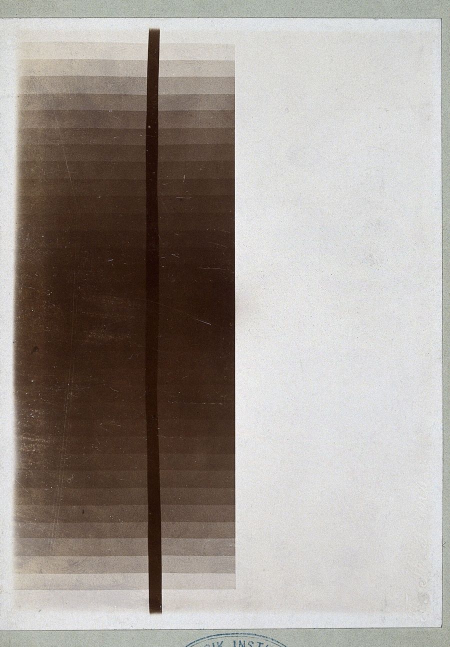
Layers of tin foil under X-rays. Photoprint from a radiograph by W. K. Röntgen, 1895. Wellcome Collection, public domain.
Soft tissue at 30 keV, from NIST’s tables: each centimetre passes about two-thirds of what reaches it.
Commit to a prediction
A centimetre of bone
Compact bone, the hard outer layer of a bone, is a little less than twice as dense as soft tissue: 1.92 grams per cubic centimetre against 1.06. A centimetre of soft tissue passes about two-thirds of these 30 keV X-rays.
How much does a centimetre of bone pass?
A forearm at 30 keV: soft tissue 6.4 cm across, with two bones, each a shell of compact bone around marrow. As on film, white is where the X-rays were stopped.
Why the first guesses made sense
A map of calcium
If you chose about half, you reasoned exactly as the rule says: twice the material, twice the slices, the fraction squared. If bone were simply denser flesh, you would be right. It is not. Soft tissue is mostly water and carbon compounds, made of light atoms: hydrogen, carbon, nitrogen and oxygen. A fifth of compact bone’s weight is calcium, and another tenth is phosphorus, in the mineral that makes bone hard.
At these energies an X-ray is stopped mainly by being swallowed whole by an atom, which throws out one of its inner electrons. That is far more likely for a heavy atom than a light one: per gram, the chance grows roughly as the cube of the atomic number. Calcium’s is 20; oxygen’s is 8. So calcium, a fifth of bone by weight, does more than two-thirds of its stopping. A radiograph is, very nearly, a map of where the calcium is.
If you chose almost nothing, you were thinking of the bright white bones on a film, and a film exaggerates. It is exposed so that flesh comes out grey, and then the difference between grey and less is shown as the difference between dark and white.
The bench
Change the X-rays
An X-ray tube’s voltage sets the energy of its X-rays. Lower energies make bone and soft tissue look more different, but more of the X-rays are stopped inside the patient, which is the dose. Higher energies pass through more easily and show less. Every radiograph is a choice between the two. The film is shown as a radiographer would expose it, so the soft tissue always comes out the same grey.
The film
A case you have not seen
A chest X-ray
A chest X-ray is taken at a much higher voltage than a hand, 120 kilovolts or more, so that the X-rays pass through the whole chest. Many of its X-rays carry between 60 and 100 keV. At 30 keV, a centimetre of bone stopped X-rays about six times as strongly as a centimetre of soft tissue.
At 100 keV, how much more strongly does bone stop them?
The same arm at 100 keV, exposed so the soft tissue is the same grey as before.
At 100 keV most X-rays that are stopped are not swallowed by atoms but knocked aside by electrons, a kind of collision called Compton scattering. Every electron does it about equally, whatever atom it belongs to. Light atoms and heavy atoms carry about the same number of electrons per gram, so at high energy a gram of bone and a gram of flesh look almost alike, and only the density is left.
That is why a chest X-ray is taken at high voltage. The ribs fade toward the grey of the flesh, the X-rays get through the whole chest with a small dose, and what stands out is the difference that remains: air in the lungs against everything else. A mammogram goes the other way, near 20 keV, because what it has to find, a small denser patch in soft tissue, needs all the contrast the low energies can give.
What you can now say
Bone stops X-rays because of its calcium, not just its density. At low energies heavy atoms swallow X-rays far more readily than light ones; at high energies every electron scatters about alike, and only density is left.
A radiograph is a map of what stopped the light, drawn with a choice of energy that trades what you can see against what the patient absorbs.
For the determined reader: the coefficients, the mixture rule, and the limits
The numbers
A narrow beam of single-energy X-rays passing a thickness L of material keeps T = exp(−(μ/ρ)ρL). NIST tabulates the mass attenuation coefficient μ/ρ for elements and for standard tissues; the densities are ICRU-44’s.
| Energy | μ soft tissue | μ compact bone | Bone / tissue | Calcium’s share of bone’s stopping |
|---|---|---|---|---|
| 20 keV | ||||
| 30 keV | ||||
| 50 keV | ||||
| 100 keV | ||||
| 150 keV |
The mixture rule
A compound’s μ/ρ is the mass-weighted sum of its elements’: μ/ρ = Σ wi(μ/ρ)i. ICRU-44 compact bone is, by mass, 3.4% hydrogen, 15.5% carbon, 4.2% nitrogen, 43.5% oxygen, 10.3% phosphorus, 22.5% calcium and traces of sodium, magnesium and sulfur. Each element’s term, divided by the total, is its share of the stopping. Photoelectric absorption per gram grows roughly as Z3/E3; Compton scattering per gram depends only on the number of electrons per gram, which is nearly the same (about half the number of nucleons) for every element except hydrogen.
Where this stops
A real tube gives a spread of energies, not one, and the spread hardens as it passes through the body, because the low energies are taken first; the “effective energies” here stand in for that spread. Scattered X-rays that still reach the film add a haze, which is why radiographers use grids. Real bones are not tubes, and real films and detectors are not linear. The physics of what stops an X-ray is exact; the arm is a sketch.
Seven weeks in Würzburg
On 8 November 1895, working alone in his laboratory at Würzburg, Röntgen was passing a current through a glass tube with most of the air pumped out, a cathode-ray tube, covered in black card so no light could escape. Across the dark room a paper screen painted with a fluorescent chemical began to glow. Whatever was reaching it came from the tube and passed through the card.
He spent seven weeks testing it almost without leaving the laboratory: through paper, wood, a thick book, sheets of metal, his own hand. He found it would darken a photographic plate, and that it went straight, without bending in a prism or a lens. He called it X, for unknown. His paper, “On a new kind of rays”, went to the Würzburg physical-medical society at the end of December. At New Year he posted copies to physicists across Europe, with prints that included his wife’s hand.
Within weeks the news was everywhere, and physicists from Manchester to New Hampshire were making their own pictures of hands and bullets in limbs. Röntgen took out no patent. He received the first Nobel Prize in Physics in 1901. The harm took longer to see: burns, then cancers, in the people who worked most closely with the tubes before anyone knew to stand back.
Where this explanation stops
Ten metres of water overhead
The air above you weighs about a kilogram for every square centimetre of ground, as much as a column of water ten metres high. X-rays see it that way. At 30 keV the whole atmosphere stops them as completely as ten metres of water would, which is why no X-ray from the sky has ever reached the ground. The X-ray sky was found only in 1962, by a small rocket that rose above the air for five minutes and saw, to everyone’s surprise, a star in Scorpius shining a thousand times brighter in X-rays than in visible light.
Every image in this exhibition was made by light that something let through: a pinhole, a lens, a filter, a window in water or in air, a hand. A radiograph makes the other half plain. It is a picture of what stopped the light.
Push the energy higher still, beyond the X-rays from a tube to the gamma rays from an atom’s nucleus, and a single grain of light carries so much energy that a detector can catch it alone, and weigh it.
If every picture is made of what was let through and what was stopped, what is a picture of?
End of chapter 11
Added to your notebook
Bone stops X-rays because of its calcium, not just its density. At high energies only density is left, which is why the X-rays are chosen for what the picture must show.
What happens when one grain of light carries enough energy to be counted on its own?
Chapter 12 · The Hardest Light